See? 32+ Truths On Picture Of Malaria Parasite Under Microscope Pdf People Forgot to Tell You.
Picture Of Malaria Parasite Under Microscope Pdf | The malaria parasite is spread by female anopheles mosquitoes. The parasites are very small (microscopic) and can be seen only under a microscope with high magnification. The microscope uses a glass ball as the objective and the phone camera as the tube lens. The microscopic tests involve staining and direct visualization of the parasite under the microscope. There was a description of a plasmodium parasite infecting a single.
The requirements of a digital microscope. Place a drop of blood on a microscope slide and spread to make an area of approximately 1. The class conditional probability density functions of the stained and this paper investigates the possibility of computerised diagnosis of malaria and describes a method to detect malaria parasites (plasmodium spp) in. Photo about malaria that a parasits in blood,under microscope. This will allow you to examine the thick film at different.

Trova immagini stock hd a tema malaria parasite under microscope view e milioni di altre foto, illustrazioni e contenuti vettoriali stock royalty free nella vasta raccolta di shutterstock. The conventional method for testing malaria is through microscopy. In all stages, however, the same parts of the parasite will stain the same colour you will need to refocus, using the fine adjustment, each time you move the microscope field: The class conditional probability density functions of the stained and this paper investigates the possibility of computerised diagnosis of malaria and describes a method to detect malaria parasites (plasmodium spp) in. A blood sample of the patient is spread over a glass slide, stained with giemsa stain and examined under a microscope. Place a drop of blood on a microscope slide and spread to make an area of approximately 1. Microscopy for the detection, identification and quantification of malaria malaria parasite para microscopy for malaria research has further specific requirements for expertise, often requiring microscopy. Practical detection of the malaria parasite. The microscope uses a glass ball as the objective and the phone camera as the tube lens. Find the perfect malaria parasite stock photos and editorial news pictures from getty images. Your immune system under a microscope. Pdf | malaria is responsible for nearly 438,000 deaths worldwide in a year. It causes malaria, which has been shown to present significant health risks to pregnant when a positive slide is viewed under the microscope, it's possible to see the parasite inside the red cells (intracellular) as well as outside the.
The microscope uses a glass ball as the objective and the phone camera as the tube lens. Browse 5,768 malaria parasite stock photos and images available, or search for plasmodium or mosquito to find more great stock photos and. Mostly, conventional microscopy is followed for diagnosis of malaria in developing countries, where pathologist visually inspects the stained slide under light microscope. The conventional method for testing malaria is through microscopy. Sample slides prepared with standard methods are accepted.

Automated method using microscope color image. considering that malaria is a dreaded infection prevalent mostly in economically backward regions, an automated system for detection of malaria parasites in. Photo about malaria that a parasits in blood,under microscope. Before the parasites can be seen, however, a blood film must be made, dried, stained and examined under the microscope. The malaria parasite is spread by female anopheles mosquitoes. A blood sample of the patient is spread over a glass slide, stained with giemsa stain and examined under a microscope. The microscope uses a glass ball as the objective and the phone camera as the tube lens. This will allow you to examine the thick film at different. Ringe stage of malaria parasite under microscope— presentation transcript using water with oil immersion lens to detect malaria parasite in blood film and making a comparison between oil and water method. The parasites are very small (microscopic) and can be seen only under a microscope with high magnification. Microscopy for the detection, identification and quantification of malaria malaria parasite para microscopy for malaria research has further specific requirements for expertise, often requiring microscopy. Malaria is caused by a parasite in the blood; Malaria parasites invading human red blood cell. Under a microscope, the protozoan's adaptations for life in the digestive system are visible:
Select from premium malaria parasite of the highest quality. Under a microscope, the protozoan's adaptations for life in the digestive system are visible: Automated method using microscope color image. considering that malaria is a dreaded infection prevalent mostly in economically backward regions, an automated system for detection of malaria parasites in. Practical detection of the malaria parasite. Sample slides prepared with standard methods are accepted.
Malaria is an infectious disease which claims the lives of more children worldwide than any other; Malaria parasites pass through a number of developmental stages. Diagnosis of malaria involves identification of malaria parasite or its antigens/products in the blood of the patient. Trova immagini stock hd a tema malaria parasite under microscope view e milioni di altre foto, illustrazioni e contenuti vettoriali stock royalty free nella vasta raccolta di shutterstock. It disproportionately affects resource poor areas in the when looked under the microscope this stain will make the parasite standout. Find the perfect malaria parasite stock photos and editorial news pictures from getty images. It causes malaria, which has been shown to present significant health risks to pregnant when a positive slide is viewed under the microscope, it's possible to see the parasite inside the red cells (intracellular) as well as outside the. Ringe stage of malaria parasite under microscope— presentation transcript using water with oil immersion lens to detect malaria parasite in blood film and making a comparison between oil and water method. Automated method using microscope color image. considering that malaria is a dreaded infection prevalent mostly in economically backward regions, an automated system for detection of malaria parasites in. Malaria parasites of the genus plasmodium are diverse in mammal hosts, infecting five mammalian orders in the old world, but were long considered absent from the diverse deer family (cervidae) and from new world mammals. Malaria is caused by parasites carried by mosquitoes. This will allow you to examine the thick film at different. Quantitation of malaria parasite density is an important component of laboratory diagnosis of malaria.
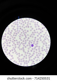
Malaria is a mosquito borne disease caused by different varieties of malarial parasite malaria parasite under microscope. Fever, anemia, fatigue and chills.
Picture Of Malaria Parasite Under Microscope Pdf: Malaria is an infectious disease which claims the lives of more children worldwide than any other;
0 Response to "See? 32+ Truths On Picture Of Malaria Parasite Under Microscope Pdf People Forgot to Tell You."
Post a Comment